 Skip to content
 Prenatal Technology Guide
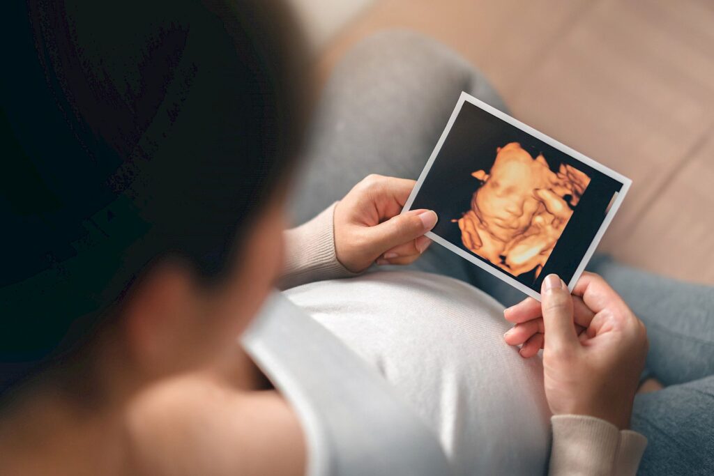
What Is 3D Ultrasound? An All-in-One Guide for Expecting Parents
Bonding with their baby before birth is one of the most cherished moments for any expectant parent. This is where 3D ultrasounds do the job! A 3D ultrasound is a modern prenatal imaging technology that produces three dimensional images of the baby when it is still in the womb. In the past, 2D ultrasound was used to see black and white images of the baby. Thanks to advanced technology, 3D ultrasound provides realistic, clear images of the baby’s body, features, and face. It is a wonderful way to bond with the parents and allow the doctors to visualize fetal growth and anomalies (if any).


How Does a 3D Ultrasound Work?
If you are wondering how 3D ultrasound technique works, we have it all mapped out for you. A 3D ultrasound provides images of the baby by combining 2D ultrasound images together from different angles. The procedure involves:
High frequency sound waves for monitoring and imagery
It is a non-invasive procedure
There is no pain for mother and the baby
It is not harmful and used across the United States
It produces clear 3D images of the baby’s features, body, and face
Why Is 3D Ultrasound Done?
3D ultrasound is conducted for both medical and non-medical reasons.
Medical Reasons
The conventionally used 2D ultrasound did not provide three dimensional picture of the baby. On the contrary, a 3D ultrasound provides a more realistic, clear, and visible pictures. It is used for monitoring of the fetal growth and any genetic anomalies. Furthermore, it also tells exactly the amniotic fluid, baby’s body, features, and contours of the body.
Non-Medical / Elective Reasons
Many parents use 3D ultrasound as a means of connecting with the baby and cherishing the comments of bonding. Furthermore, a pre-birth collection of photos can also be kept by the ultrasound. Similarly, many parents use it for gender reveal as well.
Is 3D Ultrasound Safe?
If you carry out ultrasound under the supervision of licensed professional with FDA approved machinery, it is safe and allowed all around the US.
In the USA specifically, sound waves are used for ultrasound (not radiations). There has been no risk associated with the baby or mother. It is advised to use the procedure when necessary only.
24 - 32
Weeks of Pregnancy
When Is the Best Time to Get a 3D Ultrasound?
This time period is ideal for booking your 3D ultrasound because the amniotic fluid level is adequate enough for clear images. Similarly, the baby’s facial features are also developed effectively and the womb is not too over crowded at this point. You can book your scan earlier, but it will not show detailed visibility because the body is not fully formed. Similarly, scans after 32nd week are too crowded and unclear due to the increased baby size and limited movement.
What Can You See in a 3D Ultrasound?
A 3D ultrasound can show:
- Baby’s face
- Shape of the body
- Body movements
- Facial expressions
- Nose, lips, and eyes
- Fingers and toes
- Growth and development
Image quality depends on:
- Baby’s position
- Mother’s body type
- Amniotic fluid levels
- Equipment quality
Why Is 3D Ultrasound Done?
3D ultrasound is conducted for both medical and non-medical reasons.
How Much Does a 3D Ultrasound Cost in the USA?
While the cost varies based on the clinic, in the US the range falls between:
- $150 to $300, depending on what the clinic is, its location, and reputation. Some clinics also evaluate the price based on the length of the session and whether a 4D video is included or not.
Do You Need a Doctor’s Referral for 3D Ultrasound?
- Medical 3D ultrasounds mostly call for a referral
- Elective 3D ultrasounds do not need a referral in most US states
- A medical healthcare professional to ensure the process runs smoothly.
Benefits of 3D Ultrasound
There are multiple benefits of using a 3D ultrasound, including:
- Monitoring fetal development throughout the three trimesters
- Strong emotional bonding for parents
- Early detection of anomalies
- High-quality images
Limitations of 3D Ultrasound
- It is not appropriate for diagnostic
- Image clarity depends on fetal position
- It is not a medical necessity
- Not insurance-covered if elective
Frequently Asked Questions (FAQs)
Is 3D ultrasound better than 2D?
While 2D ultrasound still remains a crucial diagnostic tool, a 3D ultrasound provides more image clarity and detection of conditions.
Can 3D ultrasound detect birth defects?
It can identify physical abnormalities of the baby before birth. However, it is important to remember that a 3D ultrasound is not similar to a diagnostic ultrasound.
Is 3D ultrasound safe for the baby?
Yes, when conducted under FDA approved conditions and within recommended limits.
Can I get a 3D ultrasound just for pictures?
Yes. Many clinics in the USA offer elective 3D/4D ultrasound services.
Difference Between 2D, 3D, 4D, and 5D Ultrasound
2D Ultrasound
A 2D ultrasound was the first ultrasound technique to exist. It produced black and white inages of the baby and was used primarily for diagnostic purposes. It was a standard tool for prenatal checkups where doctors would monitor baby’s growth, heartbeat, organs, and overall development of the body.
Key Takeaway
2D is mainly used for the medical diagnosis
3D Ultrasound
A 3D ultrasound is an advanced technique that combines different images from the 2D scan to show a more visualized 3 dimensional image of the baby. It shows baby features, body, and overall physical development. It is effective for parents bonding with the unborn child, while it is also used by doctors for medical evaluation.
Key Takeaway
3D is effective for monitoring depth and structure
4D Ultrasound
A 4D ultrasound provides realistic, moving imagery of the baby. It allows parents to cherish the moments of small baby movements like yawning, stretching, suckling thumb. It is often opted when parents want to bond with the baby or record memories for the future.
Key Takeaway
4D is usually used to check for live movements of the baby
5D Ultrasound
A 5D ultrasound is the latest technique that produces highly realistic images of the baby. The lighting, depth, and colors are all detailed and highlighted to visualize the baby effectively. It is not usually used for medical diagnosis, but it provides valuable information for seeing photo-like images of the baby
Key Takeaway
5D is opted for enhanced realism and image quality
Conclusion
A 3D ultrasound is a modern technique used to visualize the baby before birth. It is safe and effective when parents need to cherish moments of connection with the baby. It is also very emotional moment while also giving the doctors an opportunity to detect early if there is any genetic anomaly in the body.